Showing posts with label Cardiovascular System. Show all posts

Sunday, May 6, 2018

Applied anatomy of Cardiovascular System (CVS)

Applied anatomy of Cardiovascular System (CVS)


Blood Pressure


The blood pressure is the arterial pressure exerted by the blood on the arterial walls. The maximum pressure during ventricular systole is called systolic pressure; the minimum pressure during ventricular diastole is called diastolic pressure. The systolic pressure is generated by the force of contraction of the heart; the diastolic pressure is chiefly due to arteriolar tone (peripheral resistance). The heart has to pump the blood against the diastolic pressure which is a direct load on the heart. Normally, the blood pressure is roughly 120/80 mm Hg, the systolic pressure ranging from 110-130, and the diastolic pressure from 70-80. The difference between systolic and diastolic pressure is called pulse pressure.

Hemorrhage


Hemorrhage (bleeding) is the obvious result of rupture of the blood vessels. Venous hemorrhage causes oozing of blood while arterial hemorrhage causes spurting of blood

Vascular Catastrophes


Vascular catastrophes are of three types:

  1. Thrombosis
  2. Embolism
  3. Hemorrhage
All of them result in a loss of blood supply to the area of distribution of the vessel involved, unless it is compensated by collateral circulation.

Arteriosclerosis


In old age the arteries become stiff. This phenomenon it called arteriosclerosis. This causes a variable reduction in the blood supply to the tissues and a rise in systolic pressure.

Arteritis and Phlebitis


Inflammation of an artery is known as arteritis, and inflammation of a vein as phlebitis.

Atheroma


Atheroma are patchy changes developed in the tunica intima of arteries due to accumulation of cholesterol and other lipid compounds. Arteries most commonly narrowed arc those in the heart, brain, small intestine, kidneys and lower limbs. The changes are called thrombi.

Aneurysm


Aneurysm is the swelling or dilation of blood vessels where part of the wall of artery inflates like a balloon. The wall of the blood vessel at the site of aneurysm is weaker and thinner than the rest of the blood vessels. Due to its likelihood to burst it poses a serious risk to health.

Buerger’s disease 

(thromboangitis obliterans)


This is a very painful condition. There is inflammation of small peripheral arteries of the legs. The victim is a young person and a heavy smoker.

Raynaud’s phenomenon


In this condition there is spasmodic attack of pallor of the fingers due to constriction of small arteries and arterioles in response to cold.

Acute phlebothrombosis


The veins of the lower limbs are affected. Due to lack of movement of legs there is thrombus formation with mild inflammation. This thrombus may get dislodged and flow in the blood and may block any other artery. This condition is called embolism.

Varicose veins


When the vein wall is subjected to increased pressure over long time there is atrophy of muscle and elastic tissue with fibrous replacement. This leads to stretching of the vein with tortuosity and localized bulging. Venous congestion of the feet is relieved by putting feet on the stool that is higher than the trunk, helping in venous return and relief in tiredness. Varicose veins may occur at the lower end of esophagus or in the anal canal.


Functional classification of blood vessels

Functional classification of blood vessels


Functionally, the blood vessels can be classified into the following five groups.

(a) Distributing vessels, including arteries
(b) Resistance vessels, including arterioles and precapillary sphincters
(c) Exchange vessels, including capillaries, sinusoids, and postcapillary
     venules
(d) Reservoir (capacitance) vessels, including larger venules and veins 
(e) Shunts, including various types of anastomoses.


Saturday, May 5, 2018

End-Arteries

End-Arteries


Arteries which do not anastomose with their neighbors are called end arteries. Their is no collateral circulation present besides the end arteries.

Examples of end arteries


  • Central artery of retina and labyrinthine artery of internal ear are the best examples of absolute end arteries.
  • Central branches of cerebral arteries and vasa recta of mesenteric arteries.
  • Arteries of spleen, kidney, lungs and metaphyses of long bones.


End arteries 
(Vasa Recta of Mesenteric Arteries)

Importance of end arteries


Occlusion of an end-artery causes serious nutritional disturbances resulting in death of the tissue supplied by it. For example, occlusion of central artery of retina results in blindness. The results are severe because the blood flow to that region is completely stopped since there is no collateral circulation.


Anastomosis And Its Types

Anastomosis And Its Types


A precapillary or postcapillary communication between the neighboring vessels is called anastomosis. Circulation of blood through the anastomosis is called collateral circulation.

Types of Anastomosis


Anastomoses are of three types:

1 . Arterial Anastomosis


Arterial anastomosis is the communication between the arteries, or branches of arteries. It may be actual or potential.

Actual Arterial Anastomosis: In actual arterial anastomosis the arteries meet end to end. For example, palmar arches, plantar arch, circle of Willis, intestinal arcades, labial branches of facial arteries.


The Palmar Arches

Potential Arterial Anastomosis: In potential arterial anastomosis the communication takes place between the terminal arterioles. Such communications can dilate only gradually for collateral circulation. Therefore on sudden occlusion of a main artery, the anastomosis may fail to compensate the loss. The examples are seen in the coronary arteries and the cortical branches of cerebral arteries.


Coronary Anastomosis 
(A Potential Arterial Anastomosis)

2 . Venous Anastomosis


Venous anastomosis is the communication between the veins or tributaries of veins. For example, the dorsal venous arches of the hand and foot.


Dorsal Venous Arch of foot 
(Venous Anastomosis)

3 . Arteriovenous Anastomosis (Shunt)


Arteriovenous anastomosis (shunt) is the communication between an artery and a vein. It serves the function of phasic activity of the organ. When the organ is active these shunts are closed and the blood circulates through the capillaries. However, when the organ is at rest, the blood bypasses the capillary bed and is shunted back through the arteriovenous anastomosis. The shunt vessel may be straight or coiled, possesses a thick muscular coat, and is under the influence of sympathetic nervous system.


Arteriovenous Anastomosis 
(Shunt)

Shunts of simple structure are found in the skin of nose, lips and external ear. They are also common in the mucous membrane of nose and alimentary canal and the erectile tissue of sexual organs.

Preferential ‘thoroughfare channels’ are also a kind of shunts. They course through the capillary network. Many true capillaries arise as their side branches. One thoroughfare channel with its associated capillaries forms a microcirculatory unit. The size of the unit is variable from 1-2 to 20-30 true capillaries. The number of active units varies from time to time.


Sinusoids

Sinusoids


Sinusoids are small, irregular, vascular spaces which are closely surrounded by the parenchyma of the organ. They replace capillaries in certain organs, like liver, spleen, bone marrow, suprarenal glands, parathyroid glands, carotid body, etc.


Liver Sinusoids 
(Source: Eug/Wikipedia)

Characteristic Features of sinusoids


These differ from capillaries in the following respects;

  • Their lumen is wider (up to 30 micron) and irregular.
  • Their walls are thinner and may be incomplete. They are lined by endothelium in which the phagocytic cells (macrophages) are often distributed. The adventitial support is absent.
  • These may connect arteriole with venule (spleen, bone marrow), or venule with venule (liver).

Types of Circulation of Blood

Types of Circulation of Blood


In a human body, there are three types of circulation of blood:

Systemic (greater) circulation


The blood flows from the left ventricle, through various parts of the body, to the right atrium, i.e. from the left to the right side of the heart through the arteries and veins which traverse the whole body. This circulation is responsible for keeping the body tissues alive by supplying a continuous stream of blood to them.


Systemic Circulation

Pulmonary (lesser) circulation


The blood flows from the right ventricle, through the lungs, to the left atrium, i.e. from the right to the left side of the heart. This circulation is responsible for oxygenation of blood. In pulmonary circulation, the blood passes through the lungs where Carbon dioxide is eliminated and Oxygen is added to blood. In this way, the pulmonary circulation makes sure that systemic circulation remains effective.


Pulmonary Circulation

Portal circulation


It is a part of systemic circulation, which has the following characteristics.

  • The blood passes through two sets of capillaries before draining into a systemic vein.
  • The vein draining the first capillary network is known as portal vein which branches like an artery to form the second set of capillaries or sinusoids. Examples: hepatic portal circulation, hypothalamo hypophyseal portal circulation and renal portal circulation.


Portal Circulation


Components of Cardiovascular System (CVS)

Components of Cardiovascular System 

(CVS)


Cardiovascular system is the transport system of the body, through which the nutrients are conveyed to places where these are utilized, and the metabolites (waste products) are conveyed to appropriate places from where these are expelled. The conveying medium is a liquid tissue, the blood, which flows in tubular channels called blood vessels. The circulation is maintained by the central pumping organ called the heart. For Gross anatomy of the CVS, vist : Cardiovascular System: Gross Anatomy


Cardiovascular System


Cardiovascular system is a closed system of tubes made up of the following parts based on their structural and topographical characteristics.

Heart


It is a four-chambered muscular organ which pumps blood to various parts of the body. Each half of the heart has a receiving chamber called atrium, and a pumping chamber called ventricle. For detailed study of human heart, visit: Anatomy of Heart


Heart

Arteries


These are distributing channels which carry blood away from the heart.

  • They branch like trees on their way to different parts of the body.
  • The large arteries arc rich in elastic tissue, but as branching progresses there is an ever-increasing amount Of smooth muscle in their walls.
  • The minute branches which are just visible to naked eye arc called arterioles.
  • Angeion is a Greek word, meaning a vessel (blood vessel or lymph vessel). Its word derivatives are angiology, angiography, hemangioma, and thromboangitis obliterans.
For more details about arteries, visit the page: Arteries

Veins


These are draining channels which carry blood from different parts of the body back to the heart.

  • Like rivers, the veins arc formed by tributaries.
  • The small veins (venules) join together to form, larger veins, which in turn unite to form great veins called venae cavae.
For more details about veins, visit the page: Veins


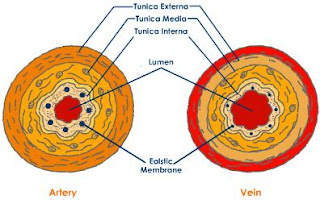
Relative Structure of Artery and Vein

For more details on the difference between arteries and veins, visit the page: Difference Between Arteries and Veins

Capillaries


These are networks of microscopic vessels which connect arterioles with the venules. These come in intimate contact with the tissues for a free exchange of nutrients and metabolites across their walls between the blood and the tissue fluid. The metabolites are partly drained by the capillaries and partly by lymphatics. Capillaries are replaced by sinusoids in certain organs, like liver and spleen.
For more details on capillaries, visit the page: Capillaries


Capillaries

Blood


Blood is a special type of tissue of human body. The character which differentiates it from rest of the tissues of human body is that it is in fluid form. It is composed of Blood Cells and Plasma. Plasma is the watery portion of blood and makes about 55% of the blood volume. The blood cells make about 45% of the blood volume and are of three types:

  • Red Blood Cells
  • White Blood Cells
  • Platelets


Bleeding Finger showing blood 
(Source: Southgeist/Wikipedia)


Capillaries

Capillaries


Capillaries (capillus = hair) are networks of microscopic endothelial tubes interposed between the metarterioles and venules. The true capillaries (without any smooth muscle cell) begin after a transition zone of 50-100 micron beyond the precapillary sphincters. The capillaries are replaced by dilated spaces in the sex organs, splenic pulp and placenta.


Capillaries

Size of Capillaries


The average diameter of a capillary is 6—8 micron, just sufficient to permit the red blood cells to pass through in ‘single file’. But the size varies from organ to organ. It is smallest in the brain and intestines, and largest in the skin and bone marrow.

Types and Structure of Capillaries


The capillaries are classified as continuous and fenestrated according to the type of junctions between the endothelial cells.

  • Continuous capillaries:
Continuous capillaries are found in the skin, connective tissue, skeletal and smooth muscles, lung and brain. These allow the small molecules to pass across their walls.

  • Fenestrated capillaries:
Fenestrated capillaries are found in the renal glomeruli, intestinal mucosa, endocrine glands and pancreas. These allow passage across their walls of larger molecules.

The capillary bed and postcapillary venules form an enormous area for the exchange of nutrients, gases, metabolites and water, between the blood and interstitial fluid. Capillaries also allow migration of leucocytes out of the vessels.


Difference between arteries and veins

Difference between arteries and veins


Arteries:


  1. Arteries carry oxygenated blood, away from the heart except pulmonary artery
  2. These are mostly deeply situated in the body
  3. These are thick-walled, highly muscular except arteries of cranium and vertebral column
  4. These posses narrow lumen
  5. Valves are absent
  6. These are reddish in color
  7. These show spurty movement of blood giving pulse
  8. Blood in arteries moves with pressure
  9. Arteries empty up at the time of death
  10. If arterial wall is injured, the blood comes out like a ‘fountain’ in a large area all around the artery

Veins:


  1. Veins carry deoxygenated blood, towards the heart except pulmonary veins
  2. These are superficial and deep in location
  3. These are thin-walled
  4. These posses wide lumen
  5. Valves are present which provide unidirectional flow of blood
  6. These are bluish in color
  7. These show sluggish movement of blood
  8. Blood in veins moves under very low pressure
  9. Veins get filled up at time of death
  10. If venous wall is injured, blood comes out, collects in a pool in a small area around vein


Veins

Veins


Veins are the blood vessels which carry the blood from peripheral tissues towards heart. They carry the deoxygenated blood, which is bluish in color and for the same reason veins appear blue.

Characteristic Features


  • Veins are thin-walled, being thinner than the arteries.
  • Their lumen is larger than that of the accompanying arteries.
  • Veins have valves which maintain the unidirectional flow of blood, even against gravity.
  • Since the venous pressure is low (7 mm Hg) the valves are of utmost value in the venous return. However, the valves are absent in:
    • The veins of less than 2 mm diameter.
    • The venae cavae.
    • The hepatic, renal, uterine, ovarian (not testicular), cerebral, spinal, pulmonary, and umbilical veins.
  • The muscular and elastic tissue content of the venous walls is much less than that of the arteries. This is directly related to the low venous pressure.
  • Large veins have dead space around them for their dilatation during increased venous return. The dead space commonly contains regional lymph nodes.


Structure of Vein

Structure of Veins


Veins are made up of usual three coats which are found in the arteries. But the coats are ill-defined, and the muscle and elastic tissue content is poor. In poorly developed tunica media, the amount of collagen fibers is more than the elastic and muscle fibers. The adventitia is thickest and best developed. The smooth muscle is altogether absent in:

  • The veins of maternal part of placenta;
  • The cranial venous sinuses and pial veins;
  • The retinal veins;
  • The veins of cancellous bone; and
  • The venous spaces of the corpora cavernosa and corpus spongiosum.

Blood and Nerve Supply of Veins


The larger veins, like the arteries, are supplied with nutrient vessels called vasa vasorum. But in the veins, the vessels may penetrate up to the intima, probably because of the low venous pressure and the low oxygen tension.
Nerves also are distributed to the veins in the same manner as to the arteries, but arc fewer in number.

Factors Helping in Venous Return


  1. Overflow from the capillaries pushed from behind by the arteries.
  2. Negative intrathoracic pressure sucks the blood into the heart from all over the body.
  3. Gravity helps venous return in the upper part of the body. .
  4. Arterial pulsations press on the venae comitantes intermittently and drive the venous blood towards the heart.
  5. Venous valves prevent any regurgitation (back flow) of the luminal blood.
  6. Muscular contractions press on the veins and form a very effective mechanism of venous return. This becomes still more effective. within the tight sleeve of the deep fascia, as is seen in the lower limbs. The calf muscles (soleus) for this reason are known as the peripheral heart. Thus the muscle pumps are important factors in the venous return.


Arteries

Arteries


Arteries are blood vessels that supply blood to the tissues of body. They carry the blood away from heart and because the blood inside them is oxygenated, they appear red in color.

Characteristic Features of Arteries


  • Arteries are thick-walled, being uniformly thicker than the accompanying veins, except for the arteries within the cranium and vertebral canal where these are thin.
  • Their lumen is smaller than that of the accompanying veins,
  • Arteries have no valves.
  • An artery is usually accompanied by veins and nerves, and the three of them together form the neurovascular bundle which is surrounded and supported by a fibro-areolar sheath.


Structure of an artery

Types and Structure of Arteries


  • Large arteries of elastic type, e.g. aorta and its main branches (brachiocephalic, common carotid, subclavian and common iliac) and the pulmonary arteries.
  • Medium and small arteries of muscular type, e.g. temporal, occipital, radial, popliteal, etc.
  • Smallest arteries of muscular type are called arterioles. They measure 50-100 micron in diameter. Arterioles divide into terminal arterioles with a diameter of 15-20 micron, and having one or two layers of smooth muscle in their walls. The side branches from terminal arterioles are called metarterioles which measure 10-15 micron at their origin and about 5 micron at their termination.
The terminal narrow end of metarteriole is surrounded by a precapillary sphincter which regulates blood flow into the capillary bed. It is important to know that the muscular arterioles arc responsible for generating peripheral resistance, and thereby for regulating the diastolic blood pressure. Microscopically, all arteries are made up of three coats.

  • The inner coat is called tunica intima.
  • The middle coat is called tunica media.
  • The outer coat is called tunica adventitia. It is strongest of all coats and merges with the perivascular sheath.
The relative thickness of the coats and the relative proportion of the muscuiar, clastic and fibrous tissues vary in different types of arteries.


Blood Supply of Arteries


The large arteries (of more than I mm diameter) are supplied with blood vessels. The nutrient vessels, called vasa vasorum, form a dense capillary network in the tunica adventitia, and supply the adventitia and the outer part of tunica media. The rest of the vessel wall (intima + inner part of media) is nourished directly by diffusion from the luminal blood. Minute veins accompanying the arteries drain the blood from the outer part of arterial wall. Lymphatics are also present in the adventitia.

Palpable Arteries


Some arteries can be palpated through the skin. These are: common carotid, facial, brachial, radial, abdominal aorta, femoral, posterior tibial and dorsalis pedis.

Nerve Supply of Arteries


The nerves supplying an artery are called nervi vascularis. The nerves arc mostly nonmyelinated sympathetic fibres which are vasoconstrictor in function. A few fibers are myelinated, and arc believed to be sensory to the outer and inner coats of the arteries. Vasodilator innervation is restricted to the following sites.

  • The skeletal muscle vessels are dilated by cholinergic sympathetic nerves.
  • The exocrine gland vessels are dilated on parasympathetic stimulation.
  • The cutaneous vessels arc dilated locally to produce the flare (redness) after an injury. The vasodilatation is produced by the afferent impulses in the cutaneous nerves which pass antidromically in their collaterals to the blood vessels (axon reflex).

Anatomy of Heart

Anatomy of Heart


Heart is a hollow muscular organ, which is somewhat pyramidal in shape. It lies within the pericardium in the mediastinum. It lies free within the pericardium except at its base where it is connected to great blood vessels.

Surfaces of Heart 


Because of its shape, the heart has three surfaces: anterior, inferior and posterior. Often the surfaces are referred to as: sternocostal (anterior), diaphragmatic (inferior) and base (posterior). The apex of the heart is directed downward, forward and to the left.

  • Anterior (Sternocostal) surface: It is formed mainly by the right atrium and right ventricle. They are separated from each other by the vertical atrioventricular groove. The right border of the anterior surface is formed by the right atrium while the left border is formed by left atrium and part of left auricle.
  • Inferior (Diaphragmatic) surface: It is formed mainly by the right and left ventricles separated by the posterior interventricular groove. The inferior surface of the right atrium into which the inferior vena cava opens, also forms part of this surface.
  • The base of the heart (posterior surface): It is formed mainly by the left atrium, into which the four pulmonary veins drain. It lies opposite to the apex. Often, the beginners think of the diaphragmatic surface of the heart as its base because of the fact that the heart rests on it, however, it should be kept in mind that the heart does not rest on its base. It rests on the diaphragmatic surface which is not the base. The posterior surface is called the base because it lies opposite to the apex of the pyramidal shaped heart.
Apex of the heart: It is formed by the left ventricle and is directed downward, forward and the left. It lies at the level of the fifth intercostals space, about 3.5 inches from the midline. The apex beat can be palpated in the region of apex of the heart.

Borders of the Heart


Because of pyramidal nature of its shape, the heart has three borders: right, left and lower. Right border is formed by the right atrium. The left border is formed by the left auricle and left ventricle. The lower border is formed by right ventricle, however, some part of it is also formed by the right atrium.

Wall of the Heart


As it was stated earlier, the heart is a hollow muscular chamber. It has strong wall that are composed of three main layers. The bulk of the wall of the heart is formed by cardiac muscles called the endocardium. On the outer side, the endocardium is covered with visceral layer of serous pericardium, known as epicardium. On the inner side, the endocardium is line with a layer of endothelium known as endocardium.

Chambers of the Heart 


Human heart is not a simple hollow pump. It has been divided by vertical septa into four chambers: two atria (right and left) and two ventricles (right and left). The atria lie superior to the ventricles. In anatomic position, the right atrium lies anterior to the left atrium and the right ventricle lies anterior to the left ventricle.

Right atrium


It consists of two regions: the main concavity and a small outpouching called auricle. At the region of junction between these two parts, on the outer side, there is a vertical groove called sulcus terminalis, which on the inner side forms a ridge known as crista terminalis. The main part of the atrium lies posterior to crista terminalis and is derived embryologically from sinus venosus. The part of the atrium, which lies in front of crista terminalis, is roughened by bundles of muscle fibers, the musculi pectinati. This anterior part is derived embryologically from primitive atrium.

Openings in the right atrium: There are four openings in the right atrium that are described below:

  • Opening for superior vena cava: It lies in the upper part and has no valves
  • Opening for inferior vena cave: It lies in the lower part and is guarder by a rudimentary, and nonfunctioning valve.
  • Opening for the coronary sinus: It lies between the opening for inferior vena cave and the atrioventricular orifice. It is also guarded by a rudimentary, nonfunctioning valve.
  • Right atrioventricular orifice: It lies anterior to the opening for inferior vena cava and is guarded by the tricuspid valve.

Right ventricle


The walls of right ventricle are much thicker as compared to those of right atrium. They show several internal projecting ridges, which are formed of muscle bundles. These ridges are known as trabeculae corneae and they give the walls a spongy appearance. They are of three types:

  • Type 1: First type of trabeculae consists of papillary muscles, which project inward. They are attached by their bases to the ventricular wall and their apices are attached by fibrous chords, known as chordae tendinae, to the cusps of the tricuspid valve.
  • Type 2: Second type consists of muscle fibers attached to the ventricular walls in the same way as the first type but they are free in the middle. One of them, known as the moderator band, crosses the entire ventricular cavity from septal to anterior wall.
  • Type 3: It is simply composed of prominent ridges.
Openings in the right ventricle: There are two openings in the right ventricle: the right atrioventricular orifice (guarded by tricuspid valve) and the opening for the pulmonary trunk (guarded by the pulmonary valve).

Tricuspid valve: It consists of three cusps each of which is formed by a fold of endocardium with a little amount of connective tissue enclosed. The bases of all three cusps are attached to the fibrous ring of the skeleton of heart and their free edges are attached to chordae tendinae. Chordae tendinae connect them to the papillary muscles, which prevent the cusps from being forced into the atrium of turning inside out during ventricular contraction.

Pulmonary valve: It guards the pulmonary orifice that leads to pulmonary trunk. It also consists of three cusps with similar formation, however, in this case the cusps are semilunar in shape. The curved lower margins and sides of each cusp are attached to the arterial wall and their open mouths are directed into the pulmonary trunk. No chordae tendinae or papillary muscles are associated with this valve. External to each cusp, the wall of pulmonary trunk bulges out to form a sinus.

Left atrium


Similar to right atrium, it consists of a main cavity and the left auricle. In anatomic position of the heart, it is situated behind the right atrium and forms greater part of the base of heart. The interior of the left atrium is smooth but the auricle possesses muscular ridges as was the case with right atrium.

Openings in the left atrium: There are a total of five openings in the left atrium, four of which are for the pulmonary veins and one is the left atrioventricular orifice. The openings of the pulmonary veins are not guarded by any valve, however, the left atrioventricular orifice is guarded by bicuspid valve.

Left ventricle


It is the strongest chamber of the heart. Its walls are three times thicker than those of the right ventricle. The reason for extra thick walls is that the left ventricle has to deal with high pressures. The pressure inside the left ventricle is about six times higher than that inside the right ventricle. In cross section, the right ventricle is circular and consequently the right ventricle is crescentic. It is because of the bulging of the interventricular septum into the right ventricle.

Openings in the left ventricle: There are two openings in the left ventricle: the left atrioventricular orifice (guarded by mitral valve, also known as bicuspid valve) and the aortic opening (guarded by aortic valve).

Mitral valve: It consists of two cusps, which have the structure similar to the cusps of tricuspid valve. The anterior of the two cusps is larger and intervenes between atrioventricular and aortic orifices. The attachment of chordae tendinae and papillary muscles is also similar to that of tricuspid valve.

Aortic valve: It is precisely similar to the pulmonary valve. Behind each cusp the aortic wall bulges to form an aortic sinus.

Relative structure of chambers of the heart


The atrial portion of heart is relatively thin walled and is divided into right and left atria by the interatrial septum. This septum runs from the anterior wall of heart backward and to the right. The ventricular portion of the heart has thick wall. It is divided into right and left ventricles by the interventricular spetum. This septum is placed obliquely and its position is indicated by anterior and posterior interventricular grooves. The lower part of the septum is thick and formed of muscular tissue while the upper part is thin and membranous.


Skeleton of Heart 


The skeleton of the heart is not an actual bony skeleton. It just consists of fibrous rings that surround right and left atrioventricular, aortic and pulmonary orifices. It is continuous with the membranous part of the interventricular septum. It separates the muscular wall of the atria from that of the ventricles and forms the basis of electrical discontinuity between them. The skeleton also supports the bases of the valve cusps and prevents them from stretching and becoming incompetent.

Conducting system of the Heart


It consists of specialized cardiac muscle present in the sinuatrial node, atrioventricular node and atrioventricular bundle along with its right and left terminal branches and Purkinje fibers (specialized cardiac muscle fibers that form the conducting system of the heart).

  • Sinuatrial node: It is located in the wall of the right atrium in the upper part of the sulcus terminalis just to the right of the opening of superior vena cava. The sinuatrial node gives origin spontaneously to rhythmical impulses that spread in all direction through the cardiac muscle of the atria. As a result, the atrial muscle contracts.
  • Atrioventricular node: It is strategically placed in the lower part of the right atrium just above the attachment of the septal cusp of the tricuspid valve. Through this node, the cardiac impulse is conducted from atria to the ventricles. The speed of conduction of impulse through the atrioventricular node is very slow, which allows sufficient time for the atria to empty their blood into the ventricles completely.
  • Atrioventricular bundle: It is also known as “the bundle of His”. It is the only pathway that connects the myocardium of the atria to the myocardium of the ventricles electrically. Thus it is the only route for transmission of impulse from atria into the ventricles. The bundle descends through the fibrous skeleton of the heart to reach the inferior border of the membranous part of the ventricular septum. When it reaches the muscular part of the septum, it divides into two branches, one for each ventricle. The right bundle branch passes to the right ventricle and the left bundle branch passes to the left. After this point, they become continuous with the fibers of Purkinje plexus.
Function of conducting system of heart: The conducting system of heart is responsible not only for generating rhythmical cardiac impulses but also for conducting these impulses rapidly throughout the myocardium of the heart. Thus it aids in coordinated and efficient contraction of different chambers of the heart.
The activities of the conducting system of heart can be influenced by the autonomic nerve supply of heart. The parasympathetic nerves slow the rhythm and diminish the speed of conduction, while the sympathetic nerves have the opposite effect.

Internodal pathways:
Impulses from the sinuatrial node have been show to travel to the atrioventricular node more rapidly than they can pass through the muscle of the heart. This can be explained by presence of specialized pathways in the atrial wall, which have a structure in between that of the Purkinje fibers and ordinary muscle cells. These specialized pathways are called internodal pathways and there are three of them in the atrial wall.

  • Anterior internodal pathway: It leaves the anterior end of the SA node and passes anterior to the superior vena cava to end in the AV node.
  • Middle internodal pathway: It leaves the posterior end of the SA node and passes posterior to the superior vena cava to end in the AV node.
  • Posterior internodal pathway: It leaves the posterior part of the SA node and descends through the crista terminalis to end in the AV node.

Arterial supply of the heart


Arterial supply of the heart consists of right and left coronary arteries. These arteries arise from the ascending aorta immediately above the aortic valve. The coronary arteries along with their major branches are distributed over the surface of the heart.

Right coronary artery: It arises from the anterior aortic sinus of the ascending aorta and runs forward between the pulmonary trunk and right auricle. After reaching the surface of the heart, it descends almost vertically into the right atrioventricular groove. After reaching the inferior border, it continues posteriorly and anastomose with the left coronary artery.

Branches of right coronary artery:

  • Right conus artery: It supplies the anterior surface of pulmonary conus and upper part of the right ventricle on anterior side.
  • Anterior ventricular branches: They are two or three in number and all of them supply the anterior surface of the right ventricle.
  • Posterior ventricular branches: They are usually two in number and both of them supply the diaphragmatic surface of the right ventricle.
  • Posterior interventricular artery: It is also known as posterior descending artery. It runs towards the apex of the heart in the posterior interventricular groove and gives of branches to the right and left ventricles. It also supplies the posterior part of the ventricular septum (not the apical part, which receives blood from the anterior interventricular branch of left coronary artery) and the atrioventricular node.
  • The atrial branches: They supply the anterior and lateral surfaces of the right atrium. They also supply the sinuatrial node. In 35% individuals, the atrial branches arise from the left coronary artery.
Left coronary artery:

It is usually larger than the right coronary artery and supplies the major part of heart. It arises from the left posterior aortic sinus of the ascending aorta and passes forward between pulmonary trunk and left auricle. Then it enters the atrioventricular groove and divides into anterior interventricular branch and circumflex branch.

Branches of left coronary artery:

  • Anterior interventricular branch: It is also known as the anterior descending branch. It runs downward in the anterior interventricular groove to the apex of the heart. After reaching the apex, it passes around to enter the posterior interventricular groove and anastomoses with the terminal branches of the right coronary artery. In 33% individuals, it ends in the apex of the heart. Anterior interventricular branch supplies the right and left ventricles along with the anterior part of the ventricular septum.
  • Circumflex artery: It is the same size as the anterior interventricular artery. It winds around the left margin of the heart in the atrioventricular groove. It is further divided into a number of branches that supply various parts of heart as described below.
    • Left marginal artery: It supplies the left margin of the left ventricle till the apex.
    • Anterior ventricular and posterior ventricular branches: They supply the left ventricle
    • Atrial branches: They supply the left atrium.

Summary of the overall arterial supply of heart in most individuals


Right coronary artery: It supplies

  • The entire right ventricle except for the small to the right
  • Variable part of the diaphragmatic surface of right ventricle
  • Posteroinferior third of the ventricular septum
  • Right atrium
  • Part of the left atrium
  • Sinuatrial node
  • Atrioventricular node
  • Atrioventricular bundle
  • A part of the Left bundle branch of atrioventricular bundle
Left coronary artery: It supplies

  • Most of the left ventricle
  • Small area of the right ventricle that is not supplied by right coronary artery
  • Anterior two thirds of the ventricular septum
  • Most of the left atrium
  • Right bundle branch
  • Left bundle branch

Venous drainage of the Heart


Most blood from the heart wall drains into the right atrium through the coronary sinus. This sinus lies in the posterior part of the anterior interventricular groove and is a continuation of the great cardiac vein. It opens into the right atrium to the left of the inferior vena cava. The small and middle cardiac veins are tributaries of the coronary sinus. The remainder of the blood is returned to right atrium through anterior cardiac vein and by other small veins that open directly into the heart chambers.

Nerve supply of the heart


The heart is innervated by sympathetic and parasympathetic fibers through the cardiac plexus situated below the arch of aorta. The sympathetic supply arises from the cervical and upper thoracic portions of the sympathetic trunk. The parasympathetic supply comes from the vagus nerve.
Sympathetic fibers, which are postganglionic, terminate on the sinuatrial node, atrioventricular node, cardiac muscle fibers, and coronary arteries. Activation of these nerves causes cardiac acceleration, increased force of contraction and dilation of coronary arteries, all of which are meant to increase blood supply to the body.
The parasympathetic fibers, which are also postganglionic, terminate on sinuatrial node, atrioventricular node and coronary arteries. Cardiac muscle fibers do not receive the parasympathetic nerve fibers. Activation of theses nerves cause reduction in rate of heart beats, and constriction of the coronary arteries. The force of contraction is not affected much by the parasympathetic activation.
Afferent fibers from the heart run with the parasympathetic nerves and the vagus nerve. Sensory impulses from the heart are carried in these afferent fibers.