Showing posts with label Nervous System. Show all posts

Sunday, May 6, 2018

Comparison of Sympathetic and Parasympathetic Nervous Systems

Comparison of Sympathetic and Parasympathetic Nervous Systems


Sympathetic and parasympathetic nervous systems are two divisions of the autonomic nervous system of body. They are closely coordinated with one another to regulate the functions of human body. In the lines below a brief comparison of the two systems is provided.

Sympathetic Nervous System


  1. All neurons forming this system originate from T1 to L2 segment of spinal cord. So it is called thoracolumbar outflow.
  2. Pre-ganglionic fibers are short, relay either in lateral ganglia or collateral ganglia
  3. Post-ganglionic fibers are long Nerve endings are adrenergic in nature except in sweat gland
  4. Functionally, sympathetic nerves are vasomotor, sudomotor and pilomotor to skin. It is seen when subject is in fear, fight and flight position. It dilates skeletal muscle blood vessels
  5. Effect is widely diffused and directed towards mobilization of resources and expenditure of energy during emergency and emotional crisis
  6. It supplies visceral blood vessels, skin. Afferents from viscera and specific area of skin reach the same spinal segment to go to the cerebrum. Since pain is better appreciated from the skin, it appears to be coming from skin rather than the viscera. This is the basis of referred pain.

Parasympathetic Nervous System


  1. All neurons forming this system originate from brain (Ill, VII, IX, X cranial nerves) and S2—S4 segment of spinal cord. So it is called craniosacral outflow.
  2. Pre-ganglionic fibers are very long reaching up to terminal ganglia mostly on viscera. Postganglionic fibers are short
  3. Nerve endings are cholinergic in nature
  4. Functionally, it is seen when subject is fully relaxed. Parasympathetic system has no effect on skin
  5. Effect is discrete, isolated, directed towards conservation and restoration of the resources of
  6. It only supplies viscera
For complete details visit the following links:


Autonomic Nervous System

Autonomic Nervous System


Autonomic nervous system is the part of the Peripheral Nervous System (PNS), which control the unconscious functions of body. Autonomic nervous system controls involuntary activities of the body, like sweating, salivation, peristalsis, etc. It differs fundamentally from the somatic nervous system in having:

  • The preganglionic fibers arising from the CNS
  • The ganglia for relay of the preganglionic fibers
  • The postganglionic fibers arising from the ganglia which supply the effectors (smooth muscles and glands).
In contrast, the somatic nerves after arising from the CNS reach their destination without any interruption.

Autonomic nervous system is divided into two more or less complementary parts: the sympathetic nervous system and parasympathetic nervous system.

The sympathetic activities are widespread and diffuse, and combat the acute emergencies. The parasympathetic activities are usually discrete and isolated, and provide a comfortable environment. Both systems function in absolute coordination and adjust the body involuntarily to the given surroundings.


Parasympathetic Nervous System

Parasympathetic Nervous System


Parasympathetic nervous system is one of the two main divisions of the autonomic nervous system.  It is responsible specifically for stimulation of activities that occur when the body is at rest. The main goal of this system is to conserve the resources of the body so that they can last longer. The parasympathetic nervous system is also known as craniosacral outflow because it arises from the brain (mixed with III, VII, IX and X cranial nerves) and sacral 2-4 segments of the spinal cord. Thus it has a cranial and a sacral part.

The preganglionic fibers are very long, reaching right up to the viscera of supply. The ganglia, called terminal ganglia, are situated mostly on the viscera and, therefore, the postganglionic fibers are very short. Parasympathetic nerve endings are cholinergic in nature, similar to the somatic nerves.

Functionally, parasympathetic activity is seen when the subject is fully relaxed. His pupils are constricted, lenses accommodated, face flushed, mouth moist, pulse slow, blood pressure low, bladder and gut contracting, and the perineal sphincters relaxed.
In general the effects of parasympathetic activity are usually discrete and isolated, and directed towards conservation and restoration of the resources of energy in the body.


Sypmathetic Nervous System

Sypmathetic Nervous System


Sympathetic nervous system is a part of the autonomic nervous system of body. It is mainly concerned with mobilization of body’s resources under stress to induce the “fight or flight response“.

Sympathetic nervous system is also known as thoracolumbar outflow because it arises from T1 to L2 segments of the spinal cord.

The myelinated preganglionic fibers (white rami communicantes) arise from the lateral column of the spinal cord, emerge through the ventral rami where the white rami are connected to the ganglia of the sympathetic chain.

Preganglionic fibers relay either in the lateral ganglia (sympathetic chain) or in the collateral ganglia, e.g. the celiac ganglion. The non-myelinated postganglionic fibers (grey rami communicantes) run for some distance before reaching the organ of supply. The adrenal medulla is a unique exception in the body; it is supplied by the preganglionic fibers.

Sympathetic nerve endings are adrenergic in nature, meaning thereby that noradrenalin is produced for neurotransmission. The only exception to this general rule is the case of cholinergic sympathetic nerves supplying the sweat glands and skeletal muscle vessels for vasodilatation.

Functionally, sympathetic nerves are vasomotor (vasocons¬trictor), sudomotor (secretomotor to sweat glands), and pilomotor (contract the arrector pili and cause erection of hair) in the skin of limbs and body wall. In addition, sympathetic activity causes dilation of pupil, pale face, dry mouth, tachycardia, rise in blood pressure, inhibition of hollow viscera, and closure of the perineal sphincters.

The blood supply to the skeletal muscles, heart and brain is markedly increased. Thus, sympathetic reactions tend to be ‘mass reactions’, widely diffused in their effect and that they are directed towards mobilization of the resources of the body for expenditure of energy in dealing with the emergencies or emotional crises (fright, fight, flight).


Myelinated and Non-myelinated Nerve Fibers

Nerve Fibers


Nerve fiber is a thread like extension of a neuron, which is formed by the axon and its covering. Thus each nerve fiber is an axon with its coverings. Larger axons are covered by a myelin sheath and are termed myelinated or medullated fibers. The fatty nature of myelin is responsible for the glistening whiteness of the peripheral nerve trunks and white matter of the CNS. Thinner axons, of less than one micron diameter, do not have the myelin sheath and are therefore termed non-myelinated or non-medullated.

However, all the fibers whether myelinated or non-myelinated have a neurolemmal sheath, which is uniformly absent in the tracts. In peripheral nerves, both the myelin and neurolemmal sheaths are derived from Schwann cells.

Myelinated Fibers


Myelinated fibers form the bulk of the somatic nerves. Structurally, they are made up of following parts from within outwards.

  1. Axis cylinder forms the central core of the fiber. It consists of axoplasm covered by axolemma.
  2. Myelin sheath, derived from Schwann cells, surrounds the axis cylinder. It is made up of alternate concentric layers of lipids and proteins formed by spiralization of the mesaxon: the lipids include cholesterol, glycolipids and phospholipids.
    Myelin sheath is interrupted at regular intervals called the nodes of Ranvier where: the adjacent Schwann cells meet. Collateral branches of the axon arise at the nodes of Ranvier. Thicker axons possess a thicker coat of myelin and longer internodes.
    Each internode is myelinated by one Schwann cell. Oblique clefts in the myelin, called incisures of Schmidt Lantermann, provide conduction channels for metabolites into the depth of the myelin and to the subjacent axon. Myelin sheath acts as an insulator for the nerve fibers.
  3. Neurolemmal sheath (sheath of Schwann) surrounds the myelin sheath. It represents the plasma membrane (basal lamina) of the Schwann cell. Beneath the membrane there lies a thin layer of cytoplasm with the nucleus of the Schwann cell. The sheaths of two cells interdigitate at the nodes of Ranvier.
  4. Neurolemmal sheath is necessary for regeneration of a damaged nerve. Tracts do not regenerate because of absence of neurolemmal sheath.
  5. Endoneurium is a delicate connective tissue sheath which surrounds the neurolemmal sheath.

Non-Myelinated Fibers


Non-myelinated fibers comprise the smaller axons of the CNS, in addition to peripheral postganglionic autonomic fibers, several types of fine sensory fibers (C fibers of skin, muscle and viscera), olfactory nerves, etc. Structurally, a non-myelinated fiber consists of a group of small axons that have invaginated separately a single Schwann cell (in series) without any spiraling of the mesaxon.
The endoneurium, instead of ensheathing individual axons, surrounds all the neurolemmal sheath by virtue of which the non-myelinated fibers, like the myelinated fibers, can regenerate after damage.


Types of Nerve Fibers

Types of Nerve Fibers


According to the size and speed of conduction, the nerve fibers are classified into three different categories, namely A, B and C. These are described in detail below:

Types of A Fibers


  1. These fibers are the thickest and fastest conducting.
  2. They are myelinated.
  3. They have a diameter of 1.5-20 micron
  4. Their speed of conduction is 4-120 m/sec, which shows that they have a really fast conduction of impulse.
  5. Examples of type A fibers are skeletomotor fibers, fusimotor fibers and afferent fibers to skin.

Types of B Fibers


  1. These fibers are medium in size, i.e. they are smaller than type A fibers but larger than type C.
  2. They are myelinated.
  3. They have a diameter of 1.5-3.5 microns.
  4. Their speed of conduction is 3-15 m/sec, which shows that they are slower than type A fibers.
  5. Examples of type B fibers are preganglionic autonomic efferents.

Types of C Fibers


  1. These fibers are the smallest and thinnest.
  2. They are non-myelinated.
  3. They have a diameter of 0.1-2 microns.
  4. Their speed of conduction is 0.5-4 m/sec, which shows that they have the slowest conduction.
  5. Examples of type C fibers are postganglionic autonomic efferents and afferent fibers to skin.


Spinal Nerves

Spinal Nerves


Spinal nerve is generally defined as a mixed nerve, which carries motor, sensory and autonomic signals between spinal cord and the body. There are 31 pairs of spinal nerves, including 8 cervical, 12 thoracic, 5 lumbar, 5 sacral, and 1 coccygeal.

Area of skin supplied by a single segment of spinal cord is called a dermatome. Each spinal nerve is connected with the spinal cord by two roots, a ventral root which is motor, and a dorsal root which is sensory.


The dorsal root is characterized by the presence of a spinal ganglion at its distal end. In the majority of nerves the ganglion lies in the intervertebral foramen.

The ventral and dorsal nerve roots unite together within the intervertebral foramen  to form the spinal nerve. The nerve emerges through the intervertebral foramen, gives off recurrent meningeal branches, and then divides immediately into a dorsal and a ventral ramus.

The dorsal ramus passes backwards and supplies the intrinsic muscles of the back, and the skin covering them.

The ventral ramus is connected with the sympathetic ganglion, and is distributed to the limb or the anterolateral body wall.

In case of a typical (thoracic) spinal nerve, the ventral ramus does not mix with neighboring rami, and gives off several muscular branches, a lateral cutaneous branch, and an anterior cutaneous branch. However, the ventral rami of other spinal nerves are plaited to form the nerve plexuses for the limbs, like the brachial plexus, lumbar plexus, etc.


Nerve Plexuses for Limbs


All nerve plexuses are formed only by the ventral rami, and never by the dorsal rami. These supply the limbs. Against each plexus the spinal cord is enlarged, e.g. ‘cervical enlargement’ for the brachial plexus, and ‘lumbar enlargement’ for the lumbosacral plexus. Plexus formation resembles a tree.

Each nerve root of the plexus (ventral ramus) divides into a ventral and dorsal division.

The ventral division supplies the flexor compartment, and the dorsal division, the extensor compartment, of the limb.

The flexor compartment has a richer nerve supply than the extensor compartment. The flexor skin is more sensitive than the extensor skin and the flexor muscles (antigravity, bulkier muscles) are more efficient and are under a more precise control than the coarse extensor muscles.

The plexus formation is a physiological or functional adaptation, and is perhaps the result of the following special features in the limbs.

1. Overlapping of dermatomes
2. Overlapping of myotomes
3. Composite nature of muscles
4. Possible migration of muscles from the trunk to the limbs
5. Linkage of the opposite groups of muscles in the spinal cord for reciprocal innervation.


Peripheral Nerves

Peripheral Nerves


The peripheral nerves are solid white cords composed of bundles (fasciculi) of nerve fibers. Each nerve fiber is an axon with its coverings. The nerve fibers are supported and bound together by connective tissue sheaths at different levels of organization of the nerve. The whole nerve trunk is ensheathed by epineurium, each fasciculus by perineurium, and each nerve fiber by a delicate endoneurium. The toughness of a nerve is due to its fibrous sheaths, otherwise the nerve tissue itself is very delicate and friable.

Blood and Nerve Supply of Peripheral Nerves


The peripheral nerves are supplied by vessels, called vasa nervorum, which form longitudinal anastomosis on the surface of the nerves. The nerves distributed to the sheaths of the nerve trunks are called nevi nervorum.


Reflex Arc

Reflex Arc


A reflex arc is the basic functional unit of the nervous system which can perform an integrated neural activity. In its simplest form, that is a monosynaptic reflex, it is made up of:

  • A receptor e.g. skin
  • A sensory or afferent neuron
  • A motor or efferent neuron
  • An effector e.g. muscle.
The complex forms of reflex arc are polysynaptic due to addition of one or more interneurons in between the afferent and efferent neurons.

An involuntary motor response of the body is called a reflex action. The stretch reflexes (tendon jerks) are the examples of monosynaptic reflexes whereas the withdrawal reflex (response to a painful stimulus) is a polysynaptic reflex.


Blood-Brain Barrier

Blood-Brain Barrier


Certain dyes, when injected intravenously, fail to stain the parenchyma of brain and spinal cord, although they pass easily into the non-nervous tissues. However, the same dyes, when injected into the ventricles, enter the brain substances easily. This indicates that a barrier exists at the capillary level between the blood and nerve cells. In simple terms, the blood brain brain barrier can be defined as the separation of the circulating blood and the brain extracellular fluid in the Central Nervous System (CNS).


Blood Brain Barrier Illustration

This means that substances present in the circulating blood cannot all pass through the blood vessels into the brain extracellular fluid. The possible structures constituting the blood-brain barrier are as follows.

(a) Capillary endothelium without fenestrations,
(b) Basement membrane of the endothelium.
(c) The end feet of astrocytes covering the capillary walls.

The barrier permits a selective passage of blood contents to the nervous tissue, and thus the toxic and harmful substances are ordinarily prevented from reaching the brain. Thus this barrier is very important for protection of brain against any harmful agents in the blood.


Glial Cells (Neuroglia)

Glial Cells (Neuroglia)


Glial Cells (neuroglia) are the non-excitable supporting cells of the nervous system. All glial cells are much smaller but far more numerous than the nerve cells. They form a major component of the nervous tissue and include the the following.


Neuroglia shown by Golgi's Method

  1. Neuroglial cells, found in the parenchyma of brain and spinal cord.
  2. Ependymal cells lining the internal cavities or ventricles;
  3. Capsular or satellite cells, surrounding neurons of the sensory and autonomic ganglia.
  4. Schwann cells, forming sheaths for axons of peripheral nerves.
  5. Several types of supporting cells, ensheathing the motor and sensory nerve terminals, and supporting the sensory epithelia.

Classification of Glial Cells


The neuroglial cells, found in the parenchyma of brain and spinal cord, are broadly classified as:

  • Macroglia, of ectodermal (neural) origin, comprising astrocytes, oligodendrocytes, and glioblasts.
  • Microglia, of mesodermal origin.


Types of neuroglia

Astrocytes


As the name suggests, these cells are star-shaped because of their numerous processes radiating in all directions. Astrocytes are of two types.

  1. Protoplasmic astrocytes, with thick and symmetrical processes are found in the grey matter.
  2. Fibrous astrocytes, with thin and asymmetrical processes, are found in the white matter.
The processes of astrocytes often end in plate-like expansions on the blood vessels, ependyma, and pial surface of the CNS.

Oligodendrocytes


As the name suggests these cells have fewer cell processes. According to their distribution, the oligodendrocytes may be intrafascicular, or perineuronal.

  • The intrafascicular cells are found in the myelinated tracts.
  • The perineuronal cells are seen on the surface of the somata of neurons.

Glioblast


These are stem cells which can differentiate into macroglial cells. They are particularly numerous beneath the ependyma.

Microglia


These are the smallest of the glial cells which have a flattened cell body with a few short, fine processes. They are often related to capillaries, and are said to be phagocytic in nature. Microglial cells are possibly derived from the circulating monocytes which migrate into the CNS during the late fetal and early postnatal life.

Functions of Glial Cells


  1. They provide mechanical support to neurons.
  2. Because of their non-conducting nature, the glial cells act as insulators between the neurons and prevent neuronal impulses from spreading in unwanted directions.
  3. They can remove the foreign material and cell debris by phagocytosis.
  4. They can repair the damaged areas of nervous tissue by proliferation (gliosis) they form glial scar tissue, and fill the gaps left by degenerated neurons.
  5. Glial cells can take up and store neurotransmitters released by the neighboring synapses. These can-either be metabolized or released again from the glial cells.
  6. They help in neuronal functions by maintaining a suitable metabolic and ionic environment for the neurons.
  7. Oligodendrocytes myelinate tracts.
  8. Ependymal cells are concerned with exchanges of materials between brain and Cerebrospinal Fluid.


Synapse

Synapse


The neurons form long chains along which the impulses are conducted in different directions. Each junction between the neurons is called a synapse. It is important to know that the contact between the neurons is by contiguity and not by continuity. The impulse is transmitted across a synapse by specific neurotransmitters, like acetylcholine, catecholamines (noradrenalin and dopamine), serotonin, histamine, glycine, GABA and certain polypeptides.


Synapse

Types of Synapses


The most common types of the synapse are axo-dendritic, somato-somatic and somato-dendritic.


In synaptic glomeruli, groups of axons make contact with the dendrites of one or more neurons for complex interactions. Functionally, a synapse may either be inhibitory or excitatory.


Difference between Axon and Dendrite

Difference between Axon and Dendrite


Both Axon and Dendrite are processes of a Neuron, however, they have some contrasting characters as described below.


Structure of a Typical Neuron 
(Source: Faigl.ladislav/Wikipedia)

Axon


  1. Only one axon is present in a neuron.
  2. It is a thin long process of uniform thickness and smooth surface.
  3. The branches of axon are fewer and at right angles to the axon.
  4. Axon contains neurofibrils and no Nissl’s granules.
  5. It forms the efferent component of the impulse.

Dendrite


  1. Dendrites are usually multiple in number in a neuron.
  2. These are short multiple processes. Their thickness diminishes as these divide repeatedly. The branches are studded with spiny projections.
  3. The dendrites branch profusely and are given off at acute angles.
  4. Dendrites contain both neurofibrils and Nissl’s granules.
  5. Dendrites form the afferent component of the impulse

Neuron

Neuron


The nervous tissue is composed of two distinct types of cells: the excitable and the non excitable. The excitable cells of the nervous system are known as neurons. They form the structural and functional unit of the nervous system and are responsible for all its activities.

Structure of a Neuron


Structure of a Typical Neuron 
(Source: Faigl.ladislav/Wikipedia)

Each nerve cell or neuron has:

  1. A cell body or perikaryon, having central nucleus and Nissl’s granules in its cytoplasm.
  2. Cell processes called neurites, which are of two types. Many short afferent processes, which are freely branching and varicose, are called dendrites.
A single long efferent process called axon, which may give off occasional branches (collaterals) and is of uniform diameter. The terminal branches of the axon are called axon terminals or telodendria.

The cell bodies (somata) of the neurons form grey matter and nuclei in the CNS (Central Nervous System), and ganglia in the PNS (Peripheral Nervous System). The cell processes (axons) form tracts in the CNS, and nerves in the PNS.

Types of Neuron


Neurons can be classified in several ways.
  • Classification according to the number of processes:

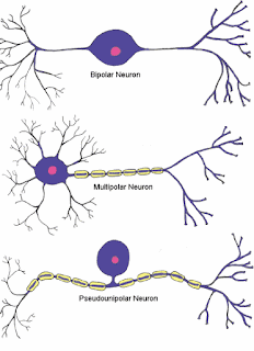
According to the number of their processes (neurites) the neurons can be divided into the following types:


Types of Neurons

  1. Unipolar neurons; e.g. mesencephalic nucleus;
  2. Pseudo-unipolar neurons, e.g. sensory ganglia or spinal ganglia
  3. Bipolar neurons, e.g. spiral and vestibular ganglia and bipolar neurons of retina.
  4. Multipolar neurons, e.g. neurons in cerebrum and cerebellum.
  • Classification according to the length of axon:

According to the length of axon, the neurons are classified as
  1. Golgi type I neurons, with a long axon
  2. Golgi type II neurons (microneurons), with a short or no axon.

Dynamic polarity of Neurons


The neurons show dynamic polarity in their processes. The impulse flows towards the soma in the dendrites, and away from the soma in the axon. However, in certain microneurons, where the axon is absent, the impulse can flow in either direction through their dendrites.


Parts of Nervous System

Nervous System


Nervous system is the chief controlling and coordinating system of the body. It controls and regulates all activities of the body, whether voluntary or involuntary, and adjusts the individual (organism) to the given surroundings. This is based on the special properties of sensitivity, conductivity and responsiveness of the nervous system.

The protoplasmic extensions of the nerve cells form the neural pathways called nerves. The nerves resemble the electricity wires. Like the electric current flowing through the wires, the impulses (sensory and motor) are conducted through the nerves.

The sensory impulses are transmitted by the sensory (afferent) nerves from the periphery (skin, mucous membranes, muscles, tendons, joints, and special sense organs) to the central nervous system (CNS). The motor impulses are transmitted by the motor (efferent) nerves from the central nervous system to the periphery (muscles and glands).

Thus the CNS is kept continuously informed about the surroundings (environment) through various sensory impulses, both general and special. The CNS in turn brings about necessary adjustment of the body by issuing appropriate orders which arc passed on as motor impulses to the muscles, vessels, viscera and glands. The adjustment of the organism to the given surroundings is the most important function of the nervous system, without which it will not be possible for the organism ta survive.


Nervous System 
(Source: TheEmirr/Wikipedia)

Parts of Nervous System


The nervous system is broadly divided into central and peripheral parts which are continuous with each other. Further subdivisions of each part are given below.

  • Central nervous system (CNS) includes:


Central Nervous System 

  1. Brain or encephalon, which occupies cranial cavity, and contains the higher governing centers.
  2. Spinal cord or spinal medulla, which occupies upper two-thirds of the vertebral canal, and contains many reflex centers.
  • Peripheral nervous system (PNS) includes:


Peripheral Nervous System 

  1. Cerebrospinal nervous system is the somatic component of the peripheral nervous system, which includes 12 pairs of cranial nerves and 31pairs of spinal nerves. It innervates the somatic structures of the head and neck, limbs and body wall, and mediates somatic sensory and motor functions.
  2. Peripheral autonomic nervous system is the visceral component of the peripheral nervous system, which includes the visceral or splanchnic nerves that are connected to the CNS through the somatic nerves. It innervates the viscera, glands, blood vessels and non-striated muscles, and mediates the visceral functions.